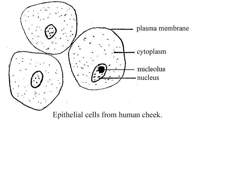
Cheek Cells Under Microscope Labeled
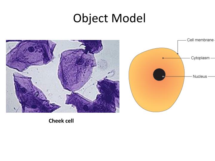
Cheek Cell
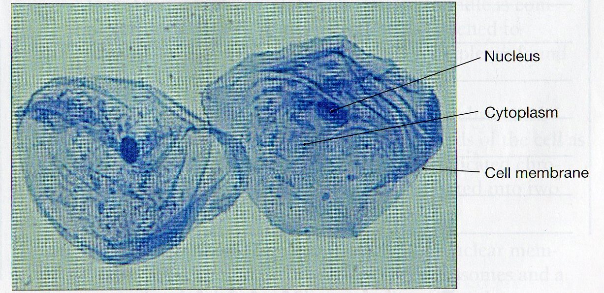
Certain stains are also used to stain specific cell structures or cell products. With cheek cells the stain methylene blue can be used. This stains the nucleus blue.
PPT Cheek cell PowerPoint Presentation, free download ID3465093
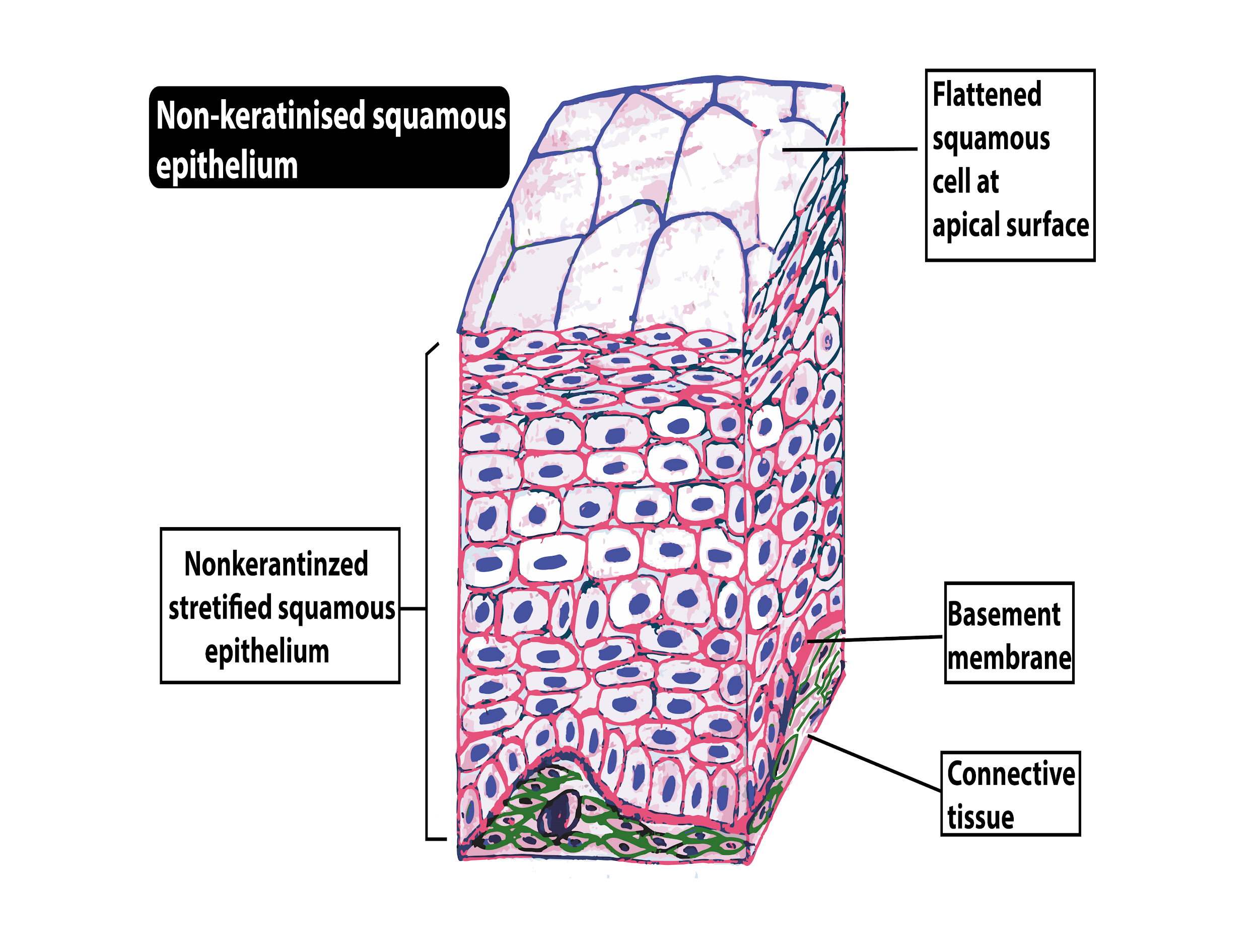
Cheek cells are epithelial cells that line the interior surface of our mouths. The base layer of cells in an epithelial structure are not actually cells, but a sticky layer on which the cells anchor. The other surface of the epithelial cell touches the outside world (like skin) or an open space (like the mouth).
Cheek Cell Diagram
1. Obtain approximately 15 mL of the 0.9% w/v saline solution to gargle with to obtain cheek cells. Swish hard and chew on your cheeks (but don't draw blood). Collect the solution in a sterile 50-mL centrifuge tube. Repeat two to three times to yield approximately 45 mL. 2. Centrifuge at 4000 rpm for 10 minutes at 4° C to pellet the cheek cells.
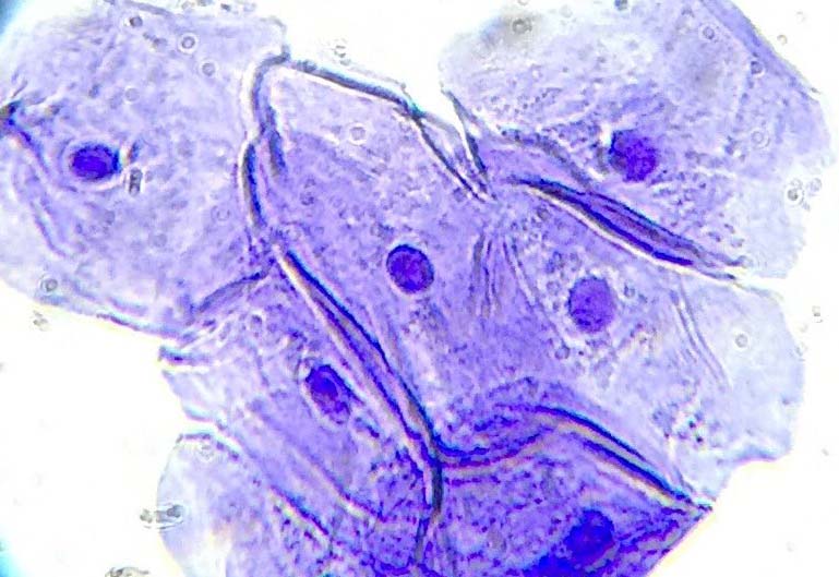
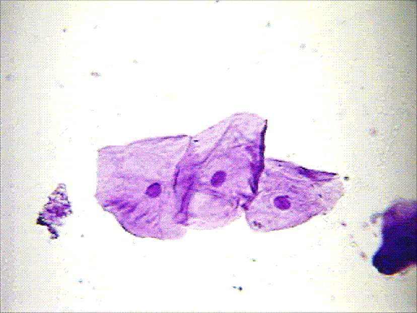
Cheek Cells
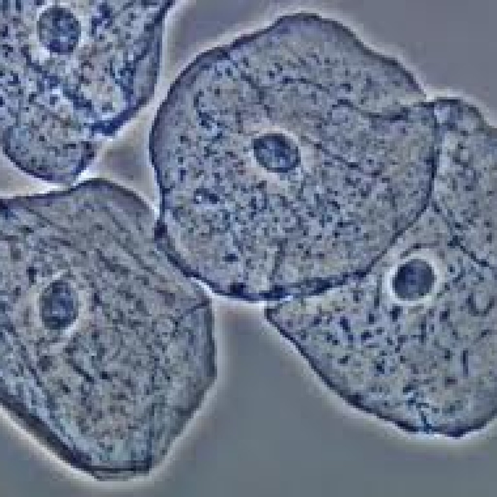
Human Cheek Epithelial Cells. The tissue that lines the inside of the mouth is known as the basal mucosa and is composed of squamous epithelial cells. These structures, commonly thought of as cheek cells, divide approximately every 24 hours and are constantly shed from the body.
Does A Cheek Cell Have A Nucleus / Cell Nucleus Contexo Info
The light microscope used in the lab is not powerful enough to view other organelles in the cheek cell. a. What parts of the cell were visible? b. List 2 organelles that were NOT visible but should have been in the cheek cell.. Fill out the Venn diagram below to show the differences and similarities between the onion cells and the Elodea cells.

Medical School • Human Cheek Cells as seen in an electron...
Hello Friends, this is my youtube channel and in this channel I used to share videos of different diagrams in easy way and step by step tutorials. These vide.
Cheek Cells Meyer Instruments
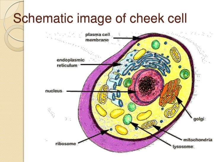
Preparing cheek cell slides to view using a light microscope is described in page 6 of this guide. Cell structure: How it is related to its function. Cytoplasm:
[DIAGRAM] Human Cheek Cell Diagram Labeled
The diagram shows the size of three organisms, different cells and other structures.. What is the width of a cheek cell compared with a Salmonella bacterium? Show answer Hide answer.
Cheek Epithelial Cells Bacteria
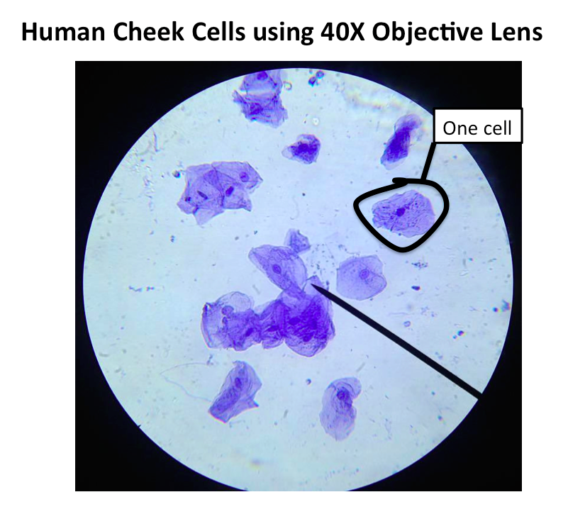
Procedure. Put a drop of methylene blue on a slide. Caution: methylene blue will stain clothes and skin. Gently scrape the inside of your cheek with the flat side of a toothpick. Scrape lightly. Stir the end of the toothpick in the stain and throw the toothpick away. Place a coverslip onto the slide. Use the SCANNING objective to focus.

Human cheek cell Diagram Quizlet
Cheek cells are readily available and easy to collect, making them an ideal and non-invasive sample for students to explore basic cell biology concepts and practice their microscopy skills. Cheek cells, fascinating eukaryotic entities, grace the lining of our mouths. These microscopic wonders boast a distinct structure that encapsulates a world.
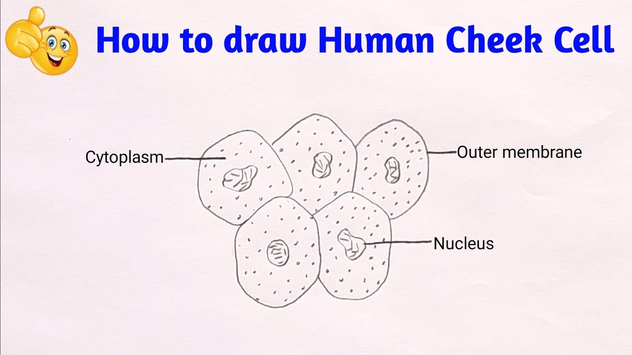
how to draw cheek cell step by step diagram of human cheek cell YouTube
5. Observe and draw an annotated diagram of your observations of a protozoan (eg. amoeba or paramecium) under your light microscope. Include any relevant information. Remember to. give your diagram a title. select only two or three cells. draw cells that are about 1⁄3 to 1⁄2 a page. state the magnification used.
Cheek Cells Under Microscope Labeled
Step 7: Observe the cheek cells under the microscope and note any differences in their shape, size or structure. Typically, cheek cells will appear as squamous epithelial cells with a flat, irregular shape. By following these steps, you can easily do a cheek smear with a microscope and look at cheek cells under a microscope.

HUMAN CHEEK CELL ( Class 8 Lesson No 8 ) YouTube
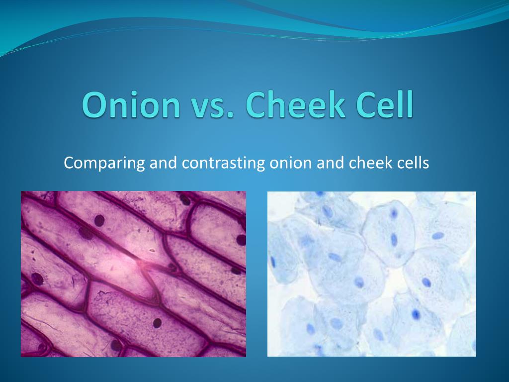
Before exploring the details of cell structure, let's understand the differences in the structure of an onion cell and a human cheek cell. Onion Cell. An onion is a multicellular (consisting of many cells) plant organism.As in all plant cells, the cell of an onion peel consists of a cell wall, cell membrane, cytoplasm, nucleus and a large vacuole.
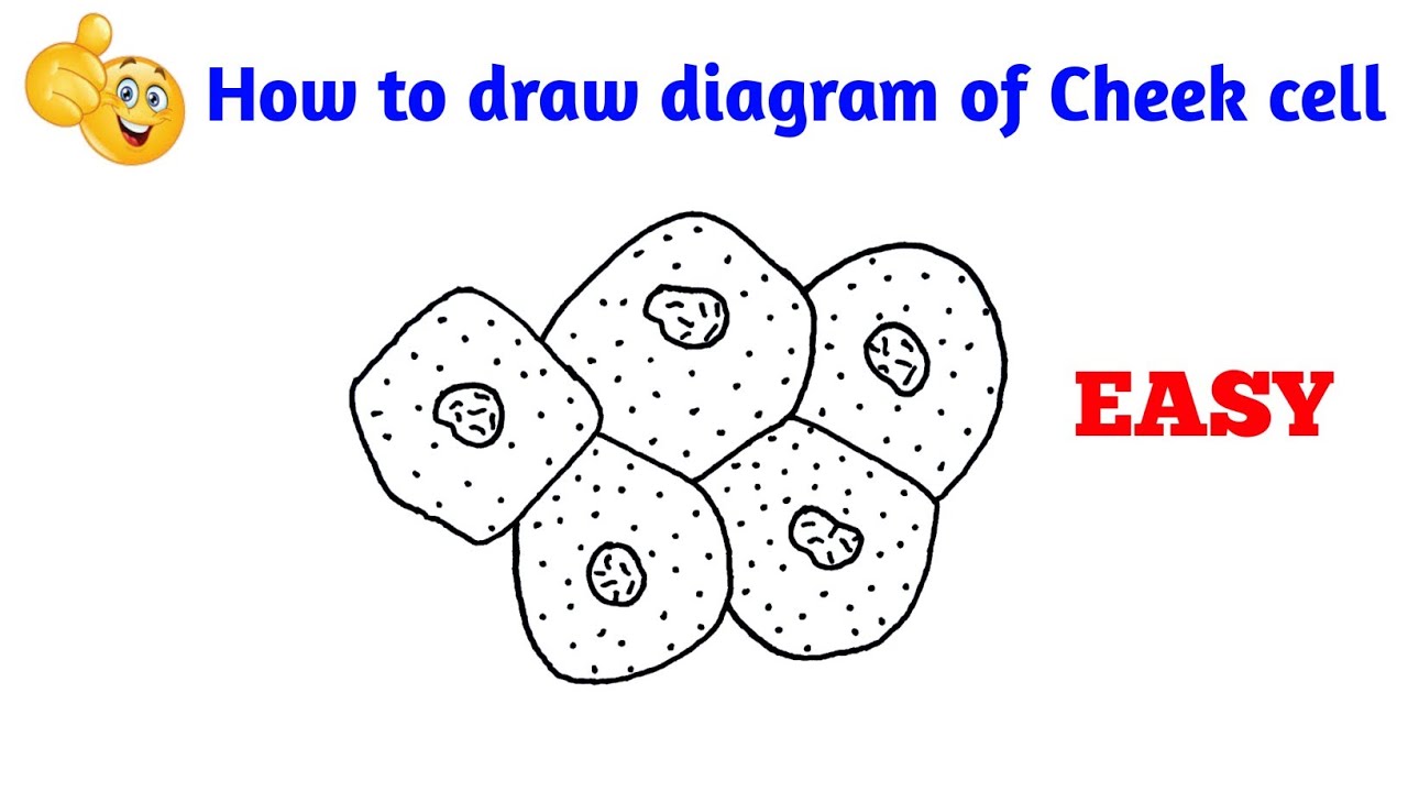
how to draw cheek cell how to draw diagram of human cheek cell YouTube
The plant cell wall is outside the cell membrane, and it provides structure for the cell. On the left is a circle representing an animal cell. The cell contains many cell parts with different shapes. A small bean-shaped cell part is labeled mitochondrion. A medium-sized circular cell part that has squiggly lines inside is labeled nucleus.

cellcheek03 Cheek Cells biologycorner Flickr
Cheek cells are eukaryotic cells (cells that contain a nucleus and other organelles within enclosed in a membrane) that are easily shed from the mouth lining. It's therefore easy to obtain them for observation.. This is an easy and fun experiment that will show kids the basic structure of a cell and its major parts. For easy identification.
Human Cheek Cell DNA extraction
The first and second branchial arches will form the bone and musculature in the cheeks. The first and second branchial pouches will development into the nerves and vessels that are within the cheek. The skin that overly the cheeks and the nerve innervation will derive from the ectoderm. The parotid gland also forms from the ectoderm layer.
